It’s a Small, Small World: Nikon Photomicrography Competition & Exhibition at Wistar |
|
January 13, 2015, Volume 61, No. 18 |
 |
 |
(Above left) Three transgenic kidneys cultured together, showing colliding branching collecting duct systems by Dr. Nils Lindstrom (16th place).
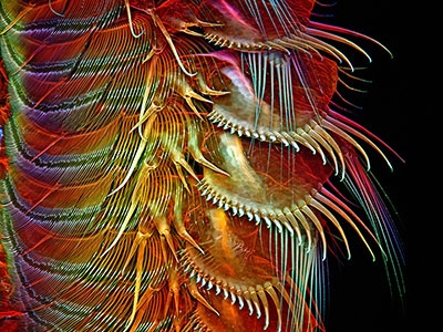
(Above right) Appendages of a common brine shrimp by Dr. Igor Robert Siwanowicz (8th Place). |
Scientific curiosity, technical expertise and pure aesthetic beauty make up the exhibition of winning images from the Nikon Small World Photomicrography Competition that arrives at The Wistar Institute with an Opening Reception on January 16, register here: www.wistar.org/NSW2015
On a daily basis, scientists peer through the microscope to see a world at cellular and sub-cellular levels in the hopes of making scientific progress. For six weeks, from January 20 through March 6, the public has the opportunity to see the best images taken through microscopes from around the world at the only venue in the Philadelphia region.
During the opening reception, guests can see video and image projections, hear a talk about the impact of microscopy, and take part in hands-on demonstrations. The Robert and Penny Fox Tower atrium feature wall, which holds a series of 15 high definition TV screens, will project a large-scale visual mix of Small World in Motion videos interspersed with Wistar research photographs. Nikon’s Eric Flem, digital imaging specialist, will discuss microscopy research as imaging technology advances offer scientists a view into new frontiers of exploration.
Also, James Hayden, previous Nikon Small World winner and managing director of Wistar’s Imaging Facility, will explain microscopy with a hands-on microscope demonstration. Hayden will discuss Wistar’s display of cell photographs along the grand staircase of the Fox Tower main entrance. Images depict various cancer cells captured by Wistar scientists to dynamically study cancer growth and cell invasion in their specific research work.
“Every day, our researchers are making discoveries through microscopy that inform our observations of cancer. Whether it’s seeing the 3-D world of our advanced tumor microenvironments, or examining fluorescently-tagged colors of live melanoma cells to help understand what treatment was more effective than another, it is done to ultimately improve our understanding of cancer to create better therapies and foster new discoveries,” said Dr. Russel E. Kaufman, Wistar president and CEO.
Celebrating 40 years, the Nikon Small World competition is a leading forum for photography from across the globe. This year’s winning images were picked from more than 1,200 entries from 79 countries and are a collection of microorganisms, cells, plants, and minerals that reveal the near-flawless techniques of microscopy (photography through microscopes), capture a decisive moment in time, and embody the scientific and artistic curiosity of the photographer. View the Nikon Small World winners, honorable mentions and images of distinction here: www.nikonsmallworld.com/galleries/photo/2014-photomicrography-competition
Related: The Images of Mongolia at the Burrison Gallery
Related: White Towers Revisited: At the Kroiz Gallery
|

